Glycocalyx of lung vascular endothelium. The world's first image capture of its three-dimensional structure
*Information related to faculty members/students and graduate schools at Gifu University here are all that of the time of interviewing.
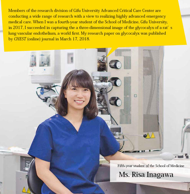
My challenge is to capture an image of the glycocalyx of the lung vascular endothelium.
Students of the School of Medicine, Gifu University, are engaged in a broad range of research projects and medical practices in laboratories of their choice from the second year to the beginning of the third year. I chose to work in the Gifu University Advanced Critical Care Center, because I thought the staff there, who have diverse medical expertise, would inspire me in my study and research. I soon found myself absorbed in my research theme, under the instruction of my supervisors. Even after my term at the center was over, I managed to find time for my research, while attending classes and continuing my club activities.
Through trial and error, I finally encountered the theme that piqued my interest the most: to capture an image of the three-dimensional structure of glycocalyx of the lung vascular endothelium.
Glycocalyx is an extremely thin coat covering the vascular endothelium. In many emergency medical cases, patients show serious symptoms of sepsis (ichorrhemia). In sepsis, white blood cells (neutrophils) release excessive protein into blood vessels to fight invading microbes, which causes glycocalyx to fall away from the vascular endothelium.
Glycocalyx plays two important roles: 1) It prevents liquid from being released outside of the blood vessels and, 2) it helps smooth blood flow. When glycocalyx falls away, liquid leaks from small holes into the blood vessel. If such a leak happens in the lungs, patients may suffer from a fatal symptom called "acute respiratory distress syndrome" (ARDS), similar to a "drowning." There are only temporary treatments for ARDS such as pumping air into the "water-filled" lung cells, or using antibacterial agents. It is therefore, imperative to visually confirm the three-dimensional structure of the glycocalyx to establish a fundamental, drastic treatments. I decided to challenge myself to capture an image of glycocalyx with an electron microscope.
"I succeeded in capturing a clear image of the glycocalyx, which could lead to medical breakthroughs in the near future. That was the moment my efforts paying paid off."
【Terminology】
Glycocalyx
Similar to the slime on the exterior of a fish,
glycocalyx is a glycoprotein and glycolipid
coating the blood vascular endothelium that
helps smooth capillary blood vessel flow.
CHEST
CHEST is a medical journal covering chest
diseases and related issues, including
pulmonology, cardiology, thoracic surgery,
transplantation, breathing, airway diseases,
and emergency medicine. Its impact factor
(IF) is 7.652 (as of 2017). In terms of
accepting and publishing research, CHEST
is considered to be one of the toughest
journals in the world today.
Sepsis
Sepsis is a life-threatening condition that
arises when the body's response to
infection causes injury to its own tissues
and organs, especially the lungs, heart,
and kidneys. It is considered a serious
case in emergency medicine.
The structure of blood vessels differs by organ, so we must observe every structure of glycocalyx accordingly by organ. There have already been successful cases of capturing images of the glycocalyx of the kidneys and heart, but because the capillary vessels surrounding both the lung tissues and layers of glycocalyx are very thin, no one had succeeded in capturing images of lung glycocalyx before me.
When I began my experiments, I realized that I needed to create test materials to shoot without damaging to the lung blood vessels. However, I learned that it was extremely difficult to permeate the lungs with fixing solution for shooting when I attempted it with mice. The glycocalyx layers always fell apart during the permeation stage. After much trial and error, as well as some kind advice from my classmates in the same laboratory, and postgraduate students at Gifu Pharmaceutical University with whom I worked, I finally found the right method of capturing the image: first, by reducing the blood pressure by dissecting the right atrial appendage; second, by slowing down the speed solution permeation; and, finally, by guiding the solution to the lungs with binding arteria carotis. My research continued until the correct method was established at every one of the above three stages. In April 2017, when I was the a fourth-year student, I finally took a picture of glycocalyx of the lung vascular endothelium. I was not certain when I saw the glycocalyx myself, but I soon felt the joy of success in seeing my supervisors celebrating my success. Later, I repeated the same experiments many times to confirm that my methods were correct, and the images of glycocalyx were authentic.
If my glycocalyx images published by "CHEST" are used by researchers and scholars to visually confirm the structure of lung glycocalyx, they may be able to prevent breakaway of glycocalyx from the blood vascular endothelium, or discover materials to detect blood vascular endothelium disorders at the earliest stage possible. My latest achievements have given me another incentive in my ongoing research efforts.
I decided to become a physician when the doctor who cared for my mother saved her and my family. After graduation, I would like to be a physician who is caring and can show compassion for both patients and their families. At the same time, I would like to build a bridge between clinical experimentation and medical treatment in the future.
microscope(SEM)
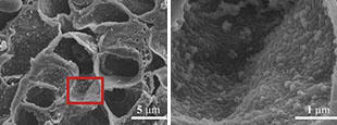
captured by SEM. The image of moss-like material on the right
is the world's first image of the glycocalyx of blood vascular
endothelium.
microscopy(TEM)
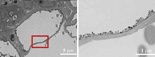
the right shows the moss-like glycocalyx covering the thin blood
vascular endothelium.
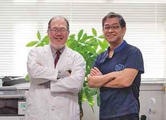
Message from Professor Shinji Ogura: "I am very pleased with her success. Her enthusiasm and tenacity led her to achieve a breakthrough."
Message from Dr. Hideshi Okada: "Like Inagawa, students can try anything without hesitation to experience the joy of discovery in the world of medicine."
Gifu University Hospital
Director of Advanced Critical Care Center
Emergency and Disaster Medicine Course, Medical Management Division,
Medical Sciences, Graduate School or Medicine
Professor Shinji Ogura (left)
Gifu University Hospital
Advanced Critical Care Center
Dr. Hideshi Okada (right)